- Information
LPIXEL Announces Launch of EIRL Chest CT
Tokyo, Japan – April 4, 2022 – LPIXEL Inc., a leader in image analysis and processing in life science and medical research, announced today that it has secured the Japanese Marketing Certification for “EIRL Chest CT”(certification number:304AGBZX00037Z00), in accordance with the Act on Securing Quality, Efficacy and Safety of Products Including Pharmaceuticals and Medical Devices (Pharmaceuticals and Medical Devices Law) and it is now commercially available.
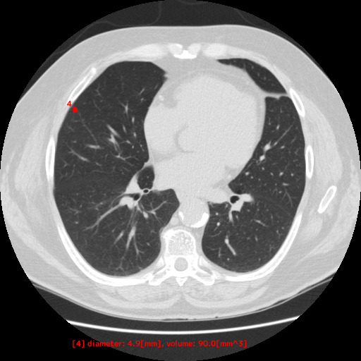
EIRL chest CT is an image analysis software that extracts regions to be observed and measures their volume and maximum diameter in the lung field of chest CT images.The software extracts “region of interest” based on CT values compared with user-set threshold in the lung field, and automatically measures their volume and maximum diameter. If the measured maximum diameter exceeds the threshold value, it is colored and displayed on the image.These functions are expected to contribute to improving the visibility of lung nodules from surrounding lung tissues when physicians read chest CT images.
Background
Lung cancer is the leading malignant neoplasm (tumor) and the leading cause of death in Japan, with a high mortality rate and number of deaths(1). To improve this situation, early detection and early treatment are essential, and chest X-ray and CT examinations are performed for early detection of lung nodules suspicious for lung cancer. In general, if any findings are found in chest X-rays performed during periodic health checkups at municipalities and business establishments, a chest CT scan is performed as a next step to reach an accurate diagnosis. In addition, chest CT scans are routinely performed at medical facilities, for example, as part of the follow-up of hospitalized patients.
The Japanese Society of CT Screening has pointed out lung cancer (pulmonary nodule) “may be overlooked even if the size exceeds 10 mm”(2). as one of the “points to keep in mind” in CT reading. Therefore, highly accurate diagnosis is required for screening chest CT examinations. However, there is a limit to the time that physicians can spend on one case due to their tight work schedule. Furthermore, it is estimated that more than 30% of medical facilities do not perform “double reading” to prevent overlooked or misdiagnosed cases(3).
LPIXEL has been developing software that assists physicians in reading and diagnosing lung nodules by extracting candidate areas as regions of interest in the lung field from chest CT images, and that measures the volume and maximum diameter of these areas.
※ About pulmonary nodules
In a chest X-ray image, air appears black, while the remaining anatomy or pathology appears white. When black areas in normal lungs turn out white with a round shape, this is called a “nodule shadow,” and lung cancer is potentially suspected. If a “nodule shadow”, one of abnormal shadows, is found on a chest X-ray or CT scan during a routine screening, it should be confirmed by a following thorough examination.
(1)Ministry of Health, Labour and Welfare, “2020 Vital Statistics Monthly Report Annual Total(https://www.mhlw.go.jp/toukei/saikin/hw/jinkou/geppo/nengai20/dl/h7.pdf)
(2)The Japanese Society of CT Screening「Criteria for Lung Nodules in Lung Cancer Screening by Low Dose CT and Concept of Follow-up Observation, 5th Edition」(https://www.jscts.org/pdf/guideline/gls5th201710.pdf)
(3)JAPAN SOCIETY OF NINGEN DOCK「Report on the status of low-dose CT lung cancer screening at member institutions of the Society」(https://www.jstage.jst.go.jp/article/ningendock/33/5/33_739/_pdf/-char/ja)
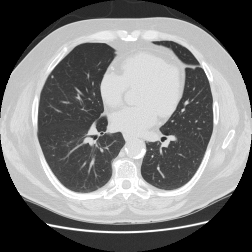
Examples
① standard dose CT
② low-dose CT
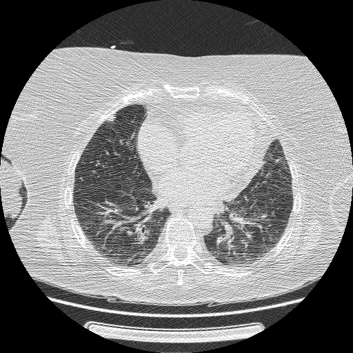
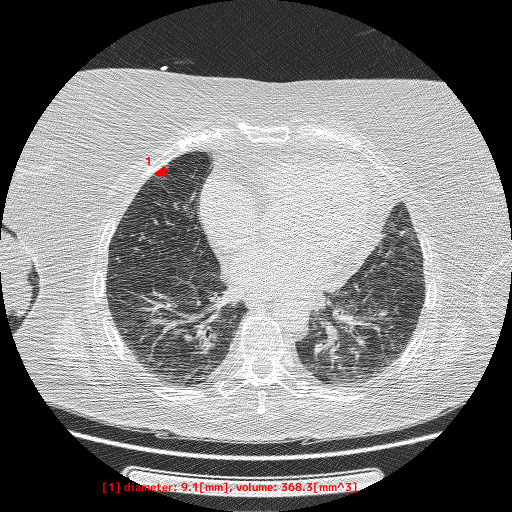
※ About Low Dose CT
Compared to conventional CT, low-dose CT can reduce the amount of radiation exposure to the examinees. On the other hand, the quality of the images on low-dose CT tends to be lower , and it is generally mentioned that reading and diagnosing is more difficult.
Contact Information about EIRL Chest CT
LPIXEL Inc. Sales Department
TEL:03-6259-1713 Email:eirl-cs@lpixel.net
URL:https://marketing.eirl.ai/ja/contact/
About EIRL
EIRL is the collective name given to LPIXEL’s AI medical image diagnostic support technology. LPIXEL aims to provide solutions that enable a faster and more accurate diagnosis by implementing its unique algorithms to analyze medical big data, such as brain MRI, chest X-rays, and colonoscopy. For more information, please visit https://eirl.ai/en/
About LPIXEL
LPIXEL is a leader in advanced image analysis and processing technology encompassing the life science field. Founded in March 2014, LPIXEL is dedicated to offering research facilities, top-tier image analysis technologies and medical diagnosis technologies, both of which adopt advanced AI technology. LPIXEL revolves around business such as its medical image analysis software, “EIRL,” and its AI-based image analysis service, “IMACEL.”
For more information, please visit https://lpixel.net/en/
Contact Information
LPIXEL Inc.
TEL:03-6259-1713 Email:pr@lpixel.net